|


|
synonyms: PRC, proximal row carpectomy
Proximal Row Carpectomy CPT
Proximal Row Carpectomy Indications
- Disabling wrist pain, secondary to SLAC wrist,
- Kienbock's disease
- Scaphoid Nonunion / AVN
- Acute/Subacute severe carpal fracture/dislocations
Proximal Row Carpectomy Contraindications
- Severe degenerative changes in the head of the capitate.
- Rheumatoid arthritis
- Patients >35y/o are more likely to require revision surgery (fusion).
Proximal Row Carpectomy Alternatives
- Wrist Arthrodesis
- Wrist arthroplasty
Proximal Row Carpectomy Planning / Special Considerations
- Radioscaphocapitate ligament is the prime stabilizer between the capitate and radius after PRC; must ensure it is preserved during surgery.
Row Carpectomy Technique
- Sign operative site.
- Pre-operative antibiotics, +/- regional block.
- General endotracheal anesthesia
- Supine position. All bony prominences well padded.
- Examination under anesthesia.
- Well padded touniquet placed high on the arm.
- Prep and drape in standard sterile fashion.
- Exsanginate the arm with Eschmar bandage, inflate tourniquet to 250mmHg.
- Oblique longitudinal incision centered on radiocarpal joint from ulnar side of radius to the base of 2nd metacarpal.
- Dissection under 2.5x/3.5x loop magnification
- Identify extensor retinaculum. Incise longitudinally, opening the 4th extensor compartment. Preserve cuff of retinaculum for later repair.
- Isolate and retract extensor tendons with penrose drain.
- Identify PINin floor of the 4th dorsal compartment and transect proximally.
- Perform T-capsulotomy centered over scapholunate joint.
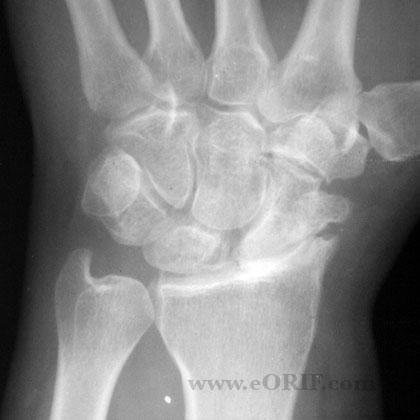
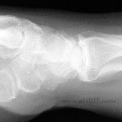
- Note dissociation of scapholunate ligament, degenerative changes in radioscaphoid joint etc, Preserved cartilage in capitate.
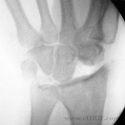
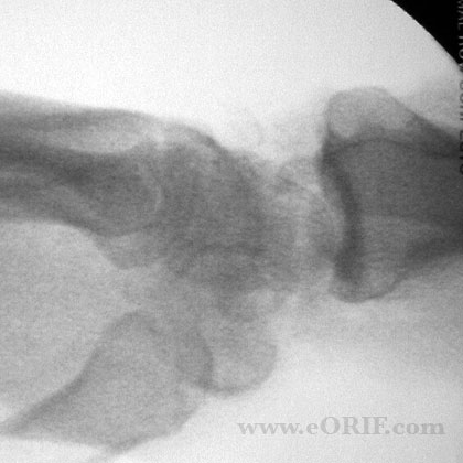
- Divide the scaphoid at its waist with in osteotome. Remove the proximal fragment.
- Excise distal fragment using a threaded Steinmann pin as a joy stick.
- Verify the integrity of the radioscaphcapitate ligament.
- Place Steinmann pin in lunate and excise lunate.
- Place Steinmann pin in triquetrum and excise.
- If needed radial styloidectomy may be performed with dissection between the 1st and 2nd dorsal compartments. Remove distal 5 to 7mm of radial styloid. Ensure radioscaphocapitate ligament is preserved.
- Irrigate.
- Repair capsule to maintain capitate in lunate fossa.
- Repair extensor retinaculum.
- Close in layers.
Proximal Row Carpectomy Complications
- Degenerative changes in the radiocapitate articulation.
- Stiffness, motion loss.
- Weakness.
- CRPS
- Continued pain.
- Instability.
- Degenerative changes in the radiocapitate articulation, Stiffness, motion loss, Weakness, CRPS Continued pain, Instability.
Proximal Row Carpectomy Follow-up care
- Post-op: Volar splint in neutral, elevation.
- 7-10 Days: Wound check, short arm cast.
- 4 Weeks: Cast removed, xray wrist. Start gentle ROM / strengthening exercises. Functional activities. Cock-up wrist splint prn / for light duty work. No heavy manual labor
- 3 Months:Full activities, may resume manual labor if adequate strength has been achieved.
- 6 Months:
- 1Yr: follow-up xrays, assess outcome
Proximal Row Carpectomy Outcomes
- >10year follow-up: 18% failure requiring fusion at an average of seven years. All in patients < 35y/o at the time of the PRC. Average flexion-extension arc = 72°, average grip strength = 91% of contralateral side. (DiDonna ML, JBJS 2004;86A:2359).
Proximal Row Carpectomy Review References
- Greens Hand Surgery
- Van Heest AE, in Masters Techniques: The Wrist, 2002
- Stern PJ, JBJS 2005;87:166
- Wyrick JD, JAAOS 2003;11:227
|


