|


|
synonyms:Scapholunate Dissociation, rotatory suplucation of the scaphoid, DISI
Scapholunate Instability ICD-10
A- initial encounter
D- subsequent encounter
S- sequela
Scapholunate Instability ICD-9
- 842.01 Sprains and strains wrist and hand; wrist; carpal (joint)
Scapholunate Instability Etiology / Epidemiology / Natural History
- Most common pattern of carpal instability
- Without treatment scapholunate advanced collapse and wrist arthritis forms.
Scapholunate Instability Anatomy
- Extrinsic wrist ligaments-extracapsular from radius to carpal or metacarpal.
- Intrinsic wrist ligaments-intracapsular between carpal bones. Thicker and stronger volarly
- Scapholunate interosseous ligament has three anatomic regions: dorsal, proximal, and palmar. Dorsal = thick and composed of short, transversely oriented collagen fibers. Proximal region = composed of fibrocartilage, with a few superficial, longitudinally oriented collagen fibers, may extend distally a few millimeters into the scapholunate joint space resembling a knee meniscus. The radioscapholunate ligament separates the proximal and palmar regions of the scapholunate interosseous ligament, often extending distally to cover the dorsal surface of the palmar region of the scapholunate interosseous ligament. Palmar = thin and composed of obliquely oriented collagen fascicles, just dorsal to and separate from the long radiolunate ligament (Berger RA, J Hand Surg 1996;21A:170)
Scapholunate Instability Clinical Evaluation
- wrist pain after a fall on the outstretched pronated hand with wrist in extension, ulnar deviation and carpal suppination.
- Vigorous hand grasp causes wrist pain.
- Decreasing repetitive grip strength.
- Watson's test: pain and a palpable clunk when the wrist is brought from ulnar deviation into radial deviation while pressure is applied to the palmar aspect of the scaphoid tubercle. (Watson HK, J Hand Surg 1988;13A:657)
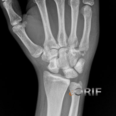
Scapholunate Instability Xray / Diagnositc Tests
- Standard views may not demonstrate scapholunatue widening. Allows consider PA Clenched first view.
- Lateral radiographs may show the scapholunate angle to be increased beyond 60°. >80° confirms SL instability. Normal scapholunate angle=47 range=30-60 degrees.
- Radiographic findings: PA radiograph, the scaphoid appears foreshortened, has a “cortical ring” sign(volar flexed scaphoid distal pole seen in cross section) and there is a scapholunate gap of greater than 3 mm. Scapholunate gap >5mm confirms scapholunate interosseous ligament disruption. Terry Thomas sign = enlarged scapholuante gap. Lunate may also appear triangular (should be quadrilateral in shape).
- PA clenched fist view in ulnar deviation accentuates widenings at the scapholunate interval.
- Comparision views of the uninjury wrist are generally indicated.
- MRI
Scapholunate Instability Classification / Treatment
Scapholunate Instability Associated Injuries / Differential Diagnosis
- Distal radius fracture
- Radial styloid fracture
- Acute carpal tunnel syndrome
- TFCC Injury
- Lunatotriquetral instability
- Lunotriquetral Coalition: a wide scapholunate gap can be a normal variant in patients with lunotriquetral coalitions.
Scapholunate Instability Complications
Scapholunate Instability Follow-up Care
Scapholunate Instability Review References
- Walsh JJ, JAAOS 2002;10:32°
|