|
|
synonyms:
Sternoclavicular Arthritis ICD-10
Sternoclavicular Arthritis ICD-9
- 715.91 Osteoarthrosis, shoulder region localized, not specified whether primary or secondary
Sternoclavicular Arthritis Etiology / Epidemiology / Natural History
- Radiographically becomes increasingly common with age. Present in 53% of patients >60years old.
- Postmenopausal women > premenopausal women & men
- Risk Factors: manual labor, radical neck disection
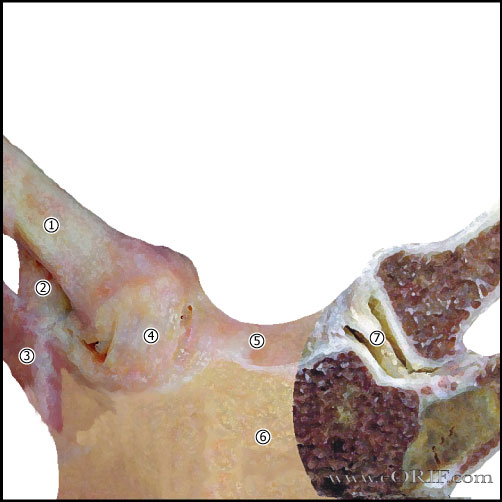
Sternoclavicular Arthritis Anatomy
- 50% of the medial head of the clavicle articulates with the sternum.
- Sternoclavicular joint allows 35° upward motion, 35° arc of A/P motion, 50° of rotation.
- Sternoclavicular joint is a diarthrodial joint.
- The only true articulation between the upper extremity and the axial skeleton.
- The medial clavicle epiphysis does not ossify until 18 to 20 y/os and does not fuse witht the medial clavicle until 23 to 25y/o. In pts whose epiphysis is unossifed it is impossible to differential between a physeal fx and a dislocation, although fx is much more likely.
- Anterior capsular ligament is the strongest of the Sternoclavicular ligaments and prevents upward displacement of the medial clavicle. (Bearn JG, J Anat 1967;101-159)
- Costoclavicular ligament (rhomboid ligament): consists of anterior and posterior faSternoclaviculariculus. Anterior faSternoclaviculariculus arises from the anteriomedial aspect of the first rib and insert more laterally on the clavicle. Posterior faSternoclaviculariculus arises lateral to the anterior faSternoclaviculariculus and inserts more medially.
- Intra-articular Disk Ligament: dense, fibrous ligament arising from the synchondral junction of the first rib to the sternum; passes through the sternoclavicular joint, dividing it into two joint spaces.
- Interclavicular ligament: arises from the upper sternum, inserts on superomedial clavicle.
- Posterior capsule most important structure in AP stability of the medial clavicle. (Spencer EE, JSES 2002;11:43)
- Safe resection length from the inferior articular surface of the medial clavicle to the most medial insertion of the costoclavicular ligament is 10 mm (Bisson LJ, JSES 2003;12:592).
Sternoclavicular Arthritis Clinical Evaluation
- Pain and swelling in the Sternoclavicular joint. Tenderness in Sternoclavicular joint.
- Increased pain with shoulder abduction and forward flexion.
- May have crepitus in the Sternoclavicular joint, or medial clavicle prominence.
- Postmenopausal women: vague aching or painless lump in Sternoclavicular joint.
Sternoclavicular Arthritis Xray / Diagnositc Tests
- AP view, apical lordotic view, serendipity view. Difficult to view on plain films. May demonstrate osteophytes.
- CT Sternoclavicularan: often helpful to demonstrate Sternoclavicularlerosis and joint space narrowing of sublte Sternoclavicular arthritis. Postmenopausal women frequently demonstrate joint subluxation.
- Labs: ESR, CRP and WBC will be normal
Sternoclavicular Arthritis Classification / Treatment
- Rest, NSAIDs, ice. Consider local corticosteriod injection.
- Surgical treatment = resection of the medial head of the clavicle. Must preserve anterior capsule & costoclavicular ligaments. (Pingsmann A, JBJS 2002;84Br:513), (Rockwood CA JR, JBJS 1997;79:387).
Sternoclavicular Arthritis Associated Injuries / Differential Diagnosis
Sternoclavicular Arthritis Complications
Sternoclavicular Arthritis Follow-up Care
Sternoclavicular Arthritis Review References
|