|


|
synonyms / keywords: Stener lesion, Gamekeepers thumb, skiers thumb, thumb UCL tear, ulnar collateral ligament of thumb,
Thumb UCL ICD-10
A- initial encounter
D- subsequent encounter
S- sequela
Thumb UCL ICD-9
- 842.12 (Sprains and strains of metacarpophalangeal joint of hand)
Thumb UCL Etiology / Epidemiology / Natural History
- Caused by rapid forceful abduction and extension at MP joint of thumb; skiers fall on abducted thumb or pole forcefully abducts thumb.
Thumb UCL Anatomy
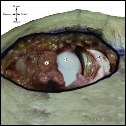
- Stener lesion = ulnar collateral ligament is displaced superficial to abductor aponeurosis. Surgical repair is indicated. (Stener B, JBJS 1962;44Br:869).
- tear of one collateral ligament removes support from that side of joint allowing phalanx to translate palmarly and rotationally.
Thumb UCL Clinical Evaluation
- tenderness ulnar side of MP joint of thumb
- proper ulnar collateral ligament is tested with thumb in 30 degrees of flexion.
- accessory ulnar collateral ligament and volar plate are tested in extension.
- increased laxity greater than 35 degrees in extention and 15 degrees in 30 degrees of flexion = complete tear.
- soft-end point indicates complete tear.
- palpable mass or Stener lesion is may be evident. This represents the palpable proximal stump of the UCL which is prevented from contacting its distal counterpart by the interposed adductor aponeurosis. In a combined cadaveric and prospective study by Heyman, laxity in excess of 35° with valgus stress testing in extension consistently indicated accessory as well as proper collateral ligament rupture. The incidence of associated Stener lesion in these cases was 89%. Conversely if at least 20° less angulation was noted with valgus stress testing in extension compared to flexion continuity of the accessory collateral ligament/palmer plate could be inferred, which prevents displacement of the proper collateral ligament and should allow ligamentous healing without operative intervention. (Heyman P, CORR 1993;292:165)
Thumb UCL Xray / Diagnositc Tests
- A/P and lateralof thumb, not hand
- evaluate for joint subluxation
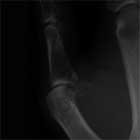
- may have associated avulsion fx
Thumb UCL Classification / Treatment
- Grade I=
- Grade II=partial tear or attenuation resulting in pain, swelling and stiffness but no significant instability. Thumb spica cast for 4 wks followed by removed splint with flexion/extension exercises until 6wks. Avoid radial directed stress such as opening jars/doors fro 10-12wks.
- Grade III=complete tears that render joint unstable= operative repair
- Bony = generally avulsion of the UCL insertion from the proximal phalangeal base. Consider arthroscopic reduction and K-wire fixation. (Badia A, Orthopedics, 2006;29:675).
- Chronic UCL injury without MCP arthritis (>6weeks from injury): primary repair with availalbe local tissue has demonstrated good long term outcomes. (Christensen, Hand (N Y). 2016 Sep; 11(3): 303–309. doi: 10.1177/1558944716628482)
- Chronic UCL injury with MCP arthritis: MCP fusion
Thumb UCL Technique
- CPT: 26540 (repair collateral ligament, MCP or IP joint)
- Video: Rossenwasser Linvatec UCL repair Using Ultrafix Minimite Suture Anchor. Ask Linvatec rep for CD if you don't have it.
- laxy S incision
- identify and preserve dorsal branches of radial sensory nerve
- identify proximal edge of adductor aponeuorisis
- incise aponeurosis longitudinally , parallel and 3-5mm palmar to extensor tendon
- evaluate joint
- repair midsubstance tear with 4-0 ethibond, avulsions repair with mini-anchors or pull-out suture(drive across with keith needle and k-wire driver)
- repair aponeurosis with 4-0 PDS
- slightly over reduce(ulnar and pronation)
- may fix joint with temporary 0.045-in K-wire
- close skin
- thumb gauntlet plaster splint
- Thumb UCL repairs have significantly reduced load to failure compared to intact ligaments (Harley BJ, J Hand Surg 2004;29Am:915).
Bony Gamekeepers ARPP
- Traction via thumb finger trap and 5lbs suspended from arm.
- Injection 1-2cc lidocaine into MCP joint.
- 1.9mm 30° scope inserted radial to the extensor tendon.
- Hematoma evacuated and synovectomy performed with 2.0mm full radius shaver.
- Fracture reduced.
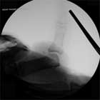
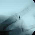
- 0.035 K-wire inserted just proximal to bony fragment, fracture reduced and fixed with K-wire under arthroscopic and fluroscopic visualization.
- K-wire cut under skin.
- Thumb-spica splint / cast for 5weeks. Pin removed under local anethesia at 5 weeks.
- (Badia A, Orthopedics, 2006;29:675).
Thumb UCL Associated Injuries / Differential Diagnosis
Thumb UCL Complications
- Stiffness
- Instability
- Neurovascular injury
- Infection
- We discussed the risks of operative intervention includeing but not limited to: incomplete relief of pain, incomplete return of function, infection, stiffness, CRPS, instability, neurovascular injury and the risks of anesthesia including heart attack, stroke and death.
Thumb UCL Follow-up Care
- 10 Days: suture removed, thumb spica cast or custom molded plastizote thumb gauntlet spint for 6 weeks. Daily active thumb MP flexion/extension exercises allowed. No pinching, grasping or resistive motions.
- 6 Weeks: wean out of thumb gauntlet splint. Begin progressive resistance exercises, occupational therapy.
- 3 Months: May return to full activities / sport if full functional strength and motion have returned. Avoid radial directed stress such as opening jars/doors for 10-12wks. Thumb gauntlet splint should be worn for any sporting activities.
Thumb UCL Review References
|


