|
synonyms:ulnar osteotomy, ulnar shortening osteotomy
Ulnar Shortening Osteotomy CPT
Ulnar Shortening Osteotomy Indications
Ulnar Shortening Osteotomy Contraindications
- Active infection
- Medically unstable patient
Ulnar Shortening Osteotomy Alternatives
- Sauve-Kapandji procedure
- Water distal ulna resection
Ulnar Shortening Osteotomy Pre-op Planning / Special Considerations
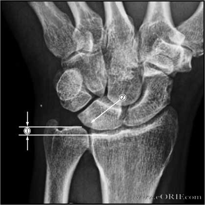
- Template amount of bone to be removed. The amount removed equals the amount of ulnar-positive variance plus the amount of ulnar-minus variance desired as measured on the neutral P/A xray. Desired unlar-minus = 2.0mm-2.5mm
- C-arm
- Ulnar shortening osteomty plate options: Synthes Small-fragment set; Acumed Osteotomy system;
Ulnar Shortening Osteotomy Technique
- Pre-operative antibiotic
- Supine position with arm table.
- Exam under anesthesia. Normal wrist ROM: Flexion= 55; Extension = 45; Radial deviation =12; Ulnar deviation =30
- Tourniquet placed high on arm.
- Prep and drape in standard sterile fashion
- Incision along subcutaneous border of ulna centered over planned osteotomy site.
- Dissection down to ulna in the interval between the ECU, FCU.
- Place 6-hole LC-DCP plate over volar ulna with the distal tip @2cm from the tip of the ulnar sytoid. Mark the center of the plate using cautery or a marker. This is the osteotomy site.
- Circumferentially subperiosteally expose the ostetomy site.
- Fix the plate to the ulna with 3.5mm screws using the distal 3 holes and standard AO-technique.
- Mark a the ulna longitudinally centered at the osteomy site to aid in restoring rotational alignment after the osteotomy.
- Remove the plate.
- Measure and mark out pre-operatively determined osteomy.
- Perform osteotomy with oscillating saw. Do not complete one of the osteotomy cuts untill the other is nearly completed. The remaining fragments become moblie after osteotomy. It is easier to make perpendicular osteotmies simultaneously. Irrigate with cool water while performing the osteotomy.
- Place plate using three distal screw holes previously made.
- Compress osteotomy site end clamp in place with reduction forceps.
- Varify appropriate variance has been restored fluoroscopically.
- Consider compressing osteotomy site with AO tensioning device.
- Fill remaining screw holes using AO technique.
- Irrigate.
- Close in layers.
- Dressing.
- Volar splint
Ulnar Shortening Osteotomy Complications
- Nonunion (@5%)
- Pain
- Stiffness
- Painful hardware
- DRUJ incongruity / arthritis
- Dorsal sensory branch of ulnar nerve injury.
Ulnar Shortening Osteotomy Follow-up care
- 7-10 follow-up: dressing changed, removable volar splint placed. Start OT for ROM.
- 6 week follow-up: check xrays. Advance therapy. Discontinue removable splint and advance to strengthening once bony union is evident.
- 12 week follow-up: check xrays
- Wrist Outcomes.
Ulnar Shortening Osteotomy Outcomes
- 77% excellent; 16% good; 3% fair; 3%poor. (Beck GK, JBJS 2005;87A:2645)
- 100% union in an average of 6–8 weeks. 72% excellent, 17% good, 11% fair. 44% required plate removal for local discomfort. (Chen NC, J Hand Surg 2003;28A:88).
- (Chun S, J Hand Surg 1993;18A:46).
Ulnar Shortening Osteotomy Review References
Green's Operative Hand Surgery: 2-Volume Set , 6e, 2010 |

