Achilles Tendon Rupture S86.019A 845.09

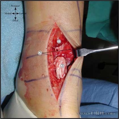
Achilles tendon rupture image

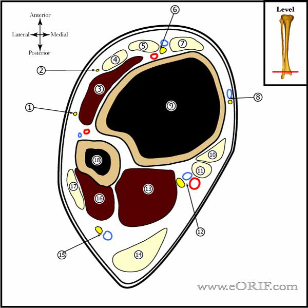
Ankle Cross section image

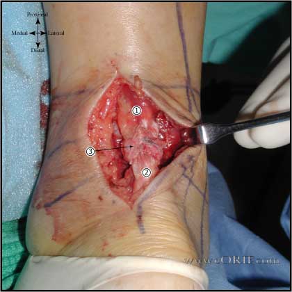
Achilles tendon rupture image

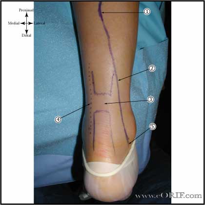
Achilles tendon rupture approach

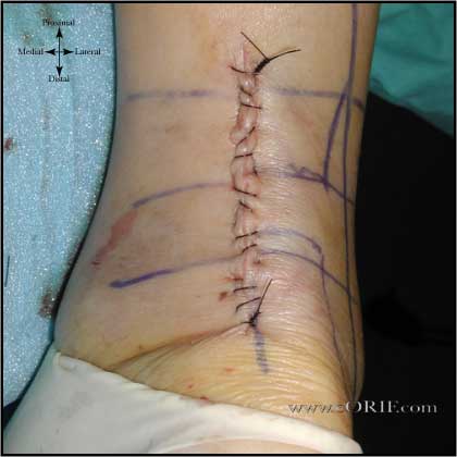
Achilles tendon incision image

synonyms: Achilles tendon tear, Achilles rupture, Achilles tear

Achilles Tendon Rupture ICD-10

Achilles Tendon Rupture ICD-9

  • 727.67 (Rupture of tendon, nontraumatic; Achilles tendon)
  • 845.09 (Sprain and strain of ankle and foot; Achilles tendon)

Achilles Tendon Etiology / Epidemiology / Natural History

  • Generally occurs in males in the 3rd to 5th decade participating in recreational sports (basketball, racket sports)
  • May be due to intrinsic degeneration or overloading (active forceful plantar flexion)
  • Has been associated with ciprofloxacin/fluoroquinolones use. (Movin T, Foot Ankle Int 1997;18:297)
  • Risk of sustaining a rupture of the contralateral side may be as high as 26% (Jessing P, Acta Chir Scand 1975;141:370)
  • Risk Factors: steriod injection into the tendon shealth or retrocalcaneal bursa, ciprofloxacin/fluoroquinolones use, type O blood, male gender, gout, hyperthyroidism, renal insufficiency, arteriosclerosis (Paavola M, Foot Ankle Clin 2002;7:501).

Achilles Tendon Anatomy

  • Achilles tendon is formed by the confluence of the Gastrocnemiusand Soleus@15cm above the calcaneous.
  • Hypovascular zone of the Achilles is 3-6cm proximal to its calcaneal insertion. (Stein V, Acta Orthop Scan 2000;71:60)
  • Sural nerve is most at risk during surgical approach.  It crosses near the midline at the level of the musculotendinous junction of the achilles (@9.8 cm from the calcaneus) before descending to its more lateral location distally. At the level of insertion of the Achilles into the calcaneus, the sural nerve is 18.8 mm from the lateral border of the Achilles tendon.   (Webb J, Foot ankle Int 2000;21:475)

Achilles Tendon Clinical Evaluation

  • Pop or snap in posterior ankle with acute pain usually associated with sudden push-off movement during sporting activity, generally in middle aged patient.
  • swollen painful ankle with posterior ecchymosis, tender posteriorly
  • Affected foot rests in slight dorsiflexion
  • Palpable defect in Achilles usually 2-4 cm above calcaneous.
  • Thompson sign = squeeze calf muscle with pt prone, feet extended off end of table.  The foot plantar flexes if tendon is intact.  No foot movement =  ruptured Achilles. (Thompson TC, J Trauma 1962:2:126).
  • O'Brien test = 25-gauge needle inserted must medial to the midline 10cm proximal to the superior border of the calcaneous. Foot is passively dorsi/plantar flexed. Needle movement in the opposite direction of the foot indicates the tendon is intact. No movement = postive test = tendon completely ruptured. Positive Thompson test with negative O'Brien test indicates parital rupture of the musculotendinous junction of the Gastrocnemius. (O'Brien T, JBJS 1984;66A:1099)
  • Hyperdorsiflexion sign: with patient prone and both knee flexed 90º, maximal passive dorsiflexion demonstrates increased dorsiflexion on injured side.
  • Repetitive heel rises: patient with a torn Achilles will not be able to perform repetitive heel rises.
  • Resting equines: ankle is normally in 7-12º of equinus when laying prone at rest. Loss of normal resting equinus position indicates Achilles rupture.

Achilles Tendon Xray / Diagnositc Tests

  • A/P, mortise and lateralankle xray indicated to r/o fracture or avulsion.  Avulsion fx best seen on lateral xray
  • MRI has been shown to be sensitive and specific, but is no better then physical exam. Disrupted tendon is best seen on T1 weighted images. 
  • Ultrasound: is useful in the diagnosis, provided skilled operator is available. Useful for determining tendon end location if considering non-op treatment. Ruptured area is hypoechogenic on ultrasound. (Hufner TM, Foot Ankle Int 2006;27:167).

Achilles Tendon Classification / Treatment

  • Initial treatment: short leg splinting in plantar flexion. Surgical and nonsurgical rx give simlar long-term outcomes. 
  • Nonsurgical treatmentrequires immobilization for 8-12 wks Disadvantages: higher recurrence rate(15.2%), higher percentage of dissatisfied patients, significant loss of power, strength and endurance compared to surgical treatment. Monitoring with Ultrasound or MRI recommended to confirm apposition of the tendon ends. Benefits: lack scar, no wound complications, less expensive. Indicated for lower demand older pt, pts on systemic steroids/methotrexate, IDDM, vascular compromise.
  • Surgical treatment. Advantages: lower re-rupture rate (1.1%), higher percentage of return to sport, greater strength, power, and endurance recovery. Disadvantages: higher cost, wound breakdown, DVT, Sural nerve injury. Indicated for: quicker return to work/sport, less atrophy, better range of motion, restore normal tension, decreased re-rupture.  (Nistor L, JBJS63A:394;1981)
  • OR= Mandelbaum BR, Am J Sports Med 1995;23:392-395
  • Percutaneous repair-risks sural nerve entrapment, decreased strength compared to open repair,: prone, zero monofilament polydioxanone suture, 2 Keith needles. Should be done within 7 days of rupture with the ankle splinted in platar flexion on the day of injury. (Bradley JP, AJSM 1990;18:188)
  • Chronic ruptures / defect >2cm-- transfer of the flexor hallucis longus (release at the knot of Henry) through the calcaneus.(Wilcox, DK Foot Ankle Int 2000;21:1004)   with concomitant V-Y advancement of the gastrocnemius. V-Y advancement can fill a defect of 2 cm to 5 cm. Flexor digitorum longus transfer, fascia lata transfer, gracilis transfer are also a viable alternative. (Wilcox DK, Foot Ankle Int. 2000 Dec;21(12):1004).
  • Achilles Avulsion from the Calcaneous: direct repair to bone with transosseous sutures or suture anchors. 

Achilles Tendon Rupture Non-operative Treatment

  • Lea RB, JBJS 1972;54A:1398
  • Weight-bearing short leg cast with the foot in a gravity equinus position for 8 weeks.
  • Cast removed at 8 weeks. Start 2.5cm heel lift and gastric stretching / strengthening.

Achilles-tendon-repair-27650

Achilles Tendon Associated Injuries / Differential Diagnosis

  • Gastrocnemious muscle tear

Achilles Tendon Complications

  • Skin necrosis
  • Infection / Dehiscence
  • Painful scar tissue
  • DVT
  • Sural nerve injury / neuroma

Achilles Tendon Follow-up Care

  • Post-Op: place in a gravity equinus splint, non-weight bearing
  • 10 day f/u, casted at 20º flexion, NWB; Consider functional boot with heel wedge. Consider early weight bearing if repair and tissues are adequate.
  • 4wks; functional boot with 20º heel wedge. WBAT. Begin gentle passive ROM, no passive dorsi flexion.
  • 6-8wks; 1” heel lift, begin active physical therapy:passive stretching, active theraband. Heel lift continued until able to reach 10 degrees of dorsiflexion.
  • Perform stretching exercises prior to commencing any sports permanently. May return to sports at 3-4 months.
  • Achilles Tendon Rehab Protocol

Achilles Tendon Review References

  1. DeLee & Drez's, Orthopaedic Sports Medicine: 3e;  2009
  2. Calhoun JH, in Masters Technique Foot and Ankle, 2nd ed, 2002
  3. Saltzman CL, JAAOS, 1998;6:316
  4. Wilcox DK, Bohay DR, Anderson JG. Treatment of chronic Achilles tendon disorders with flexor hallucis longus tendon transfer/augmentation. Foot Ankle Int. 2000 Dec;21(12):1004-10.
  5. Den Hartog BD. Flexor hallucis longus transfer for chronic Achilles tendonosis. Foot Ankle Int. 2003 Mar;24(3):233-7
  6. Myerson MS. Achilles tendon ruptures. Instr Course Lect. 1999;48:219-30