|


|
synonyms: capsular release, adhesive capsuliitis release, frozen shoulder release
Arthroscopic Capsular Release CPT
Arthroscopic Capsular Release Indications
- Chronic painful Posterior Capsular Contracture which has failed to respond to posterior capsular stretching.
- Post traumatic of post surgical Adhesive Capsulitis.
- Idiopathic Adhesive Capsulitis which has failed to respond to prolonged non-operative treatment (frequent night pain awaking patient from sleep).
Arthroscopic Capsular Release Contraindications
- Acute/mild Idiopathic Adhesive Capsulitis.
- Comorbidities preventing anesthesia.
Arthroscopic Capsular Release Alternatives
- Physical therapy.
- Supervised neglect (Diercks, RL. JSES 13:499; 2004).
- Open capsular release (Omari A, JSES 2001;10:353).
Arthroscopic Capsular Release Pre-op Planning / Special Considerations
- Technique and releases depend on underlying cause of pre-operative motion limits.
- Ensure a hook-tip radiofrequency device (Vapr Hook Tip, DePuyMitek) is available.
Arthroscopic Capsular Release Technique
- Pre-operative antibiotics, +/- regional block. Consider continuous interscalene anethesia (infusion of 0.25% bupivacaine at a rate of 6 mL per hour administered for 48 hours postop. (Warner JJ, JBJS 1997A;79:1151).
- General endotracheal anesthesia
- Beach chair, or lateral position. All bony prominences well padded.
- Examination under anesthesia.
- Prep and drape in standard sterile fashion.
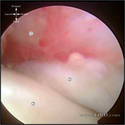
- See Shoulder Arthroscopy. Note synovial villi in the rotator interval, petechial hemorrhaging, and thickened scared capsule indicative of Adhesive Capsulitis. It is generally impossible to view inferiorly initially due to contracture.
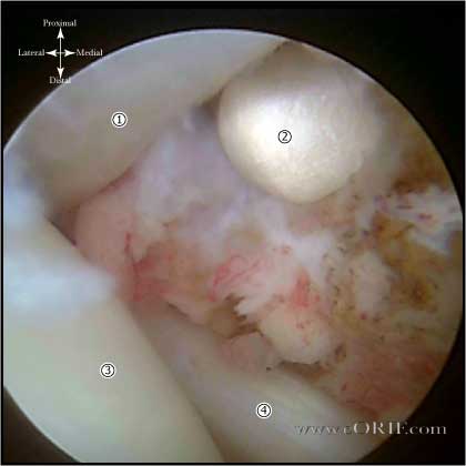
- Use cautery to release the middle glenohumeral ligament and excise entire rotator interval until coracoid process is well visualized. The vessel of the rotator interval will need to be coagulated.
- Arthroscope placed in anterosuperior portal to visualize the posterior capsule.
- The posterior capsule is generally thickened and shortened in patients with posterior capsular contracture.
- Electrocautery device is placed through the posterior portal.
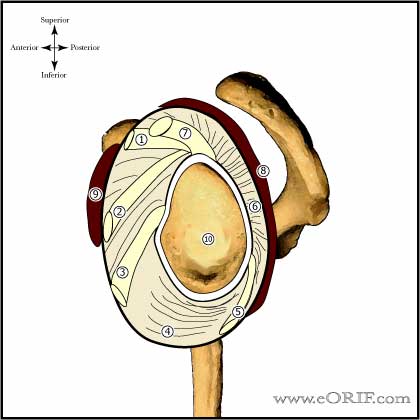
- Divide posterior capsule, adjacent to the glenoid, rim beginning just posterior to the biceps tendon origin on the superior glenoid rim(11 o’clock position), continuing inferiorly to the 8 o’clock position.
- Division of the capsule can be judged by visualizing the muscle fibers of the rotator cuff after division.
- Debride edges with an arthroscopic shaver to create a wider gap in the resected capsule to help avoid recurrence.
- Release past the 8 o'clock postion risks injury to the axillary nerve
- Remove scope and perform gentle manipulation to complete the release of any remaining capsular fibers and verify internal rotation has been restored.
- Selective Posteroinferior Capsulotomy (throwing athlete): capsulotomy is made 0.25 inches away from the labrum from the 9 o’clock position to the 6 o’clock position. Capsule is often 6 mm thick. (Burkhart SS, Arthroscopy 2003;19:404).
- Consider subacromial bursectomy or subacromial decompression. (Ticker JB, Arthroscopy 2000;16:27).
- Rotator interval is release from medial to lateral using electocautery or radiofrequency
- Release capsule lateral to labrum from superior to inferior including release of the middle GH ligament and anterior inferior band of inferior GH ligament. (improves ER). A hook-tip radiofrequency device (Vapr Hook Tip, DePuyMitek) is best used for capsular releases.
- Release inferiorly just lateral to labrum including posterior band of inferior GH ligament. (important for forward elevation and IR) Axillary nerve lies on the capsule in the lateral half of the axillary recess, ensure release is as far medial as possible to ensure nerve is not injured. Arm positioned in abduction and external rotation decreases risk of axillary nerve injury.
Arthroscopic Capsular Release Complications
- Proximal humerus fracture
- Instability
- Stiffness / Arthrofibrosis
- Chondral Injury / Arthritis
- Infection
- RTC tear
- NVI (Axillary nerve palsy)
- Fluid Extravastion / Compartment Syndrome
- Complex Regional Pain Syndrome
- Synovial fistula
- Hemarthrosis / hematoma
- DVT / PE
Arthroscopic Capsular Release Follow-up care
- Passive motion with both morning and afternoon therapy starting on post-op day 1. Begin self-assisted motion exercises ASAP.
- Daily supervised therapy 5 days per week in addition to a home-exercise program including pulleys and cane assisted motion in all planes.
- Gradually taper to home exercise program over 4-8 weeks.
- Patients are encouraged to to use the arm for daily activites ASAP. Swimming is encouraged to assist in ROM.
Arthroscopic Capsular Release Outcomes
- Internal rotation in adduction has been shown to improve a mean of four spinous-process levels and 42° in abduction. (Warner JJ, JBJS 1997A;79:1151).
- (Diwan DB, Murrell GA, Arthroscopy, 2005;21:1105).
Arthroscopic Capsular Release Review References
- Bach HG, JAAOS 2006 14: 265
- 30°
|


