|


|
synonyms:
Femoral Neck Fracture CPT
Femoral Neck Fracture Cannulated Screws Indications
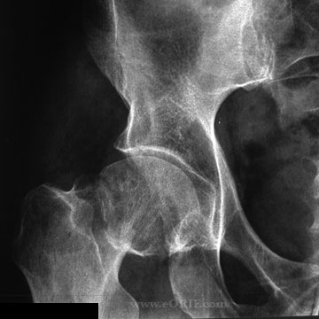
- Nondisplaced femoral neck fracture
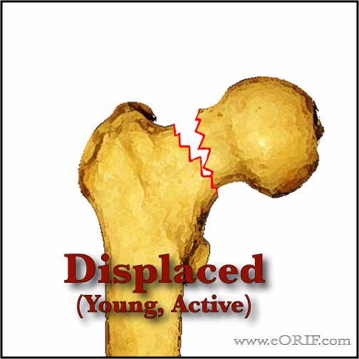
- Displaced femoral neck fracture in young, active patient
Femoral Neck Fracture Cannulated Screws Contraindications
- Displaced femoral neck fracture in elderly, inactive patient
- Rheumatoid arthritis
- Moderate osteoarthritis
- Poor bone density
- Limited life expectancy
- Pathologic fracture.
Femoral Neck Fracture Cannulated Screws Alternatives
- Hemiarthroplasty
- THA
- Dynamic hip screw with derototation screws
Femoral Neck Fracture Cannulated Screws Pre-op Planning
- Surgery should be performed ASAP after medical clearance, usually within 24-48 hours. Medical conditions should be stabilized before surgery. (Sexson SB, JOT 1988;1:298)
- Anestesia: type has not been shown to affect outcomes. (Davis FM, Br J Anaesth 1987;59:1080)
- Acceptable reduction = 15 degrees of valgus angulation, <10 degrees of anterior or posterior angulation (Koval KJ, JAAOS 1994;2:141).
Femoral Neck Fracture Cannulated Screws Technique
- Sign operative site.
- Pre-operative antibiotics, +/- regional block.
- General endotracheal anesthesia vs epidural vs spinal
- Supine on fracture table. All bony prominences well padded. All bony prominences well padded.
- Ensure adequacy of reduction with fluoroscopy before prepping and drapping. Reduction usually obtained with gentle traction and internal rotation.
- Prep and drape in standard sterile fashion. Usually with shower curtain.
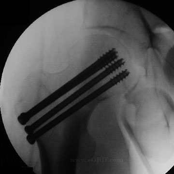
- If anatomic reduction can be achieved three parallel cannulated screws are placed. See specific manufacture technique to the right for cannulated screw techniques.
- Screws should be placed proximal to the lesser trochanter to avoid post-operative subtrochanteric femur fracture.
- Staight lateral incision starting just distal to greater trochanter and expended distally.
- Deep fascia divided in line with skin incision. Interval between tensor fascia and gluteus medius muscles is developed.
- Anterior capsule is identify by elevating pericapsular fat with a Hohman retractor.
- Capsule open sharply along the femoral neck. Evacuate hematoma.
- Fracture is reduced anatomically and reduction is varified fluoroscopically. Consider using a Schanz pain in the prosimal lateral femur distal to the site of screw placement is reduction is difficult.
- Three parallel cannulated screws are placed. There is no benefit in using >3 screws. (Springer ER, CORR 1991;267:85).
- Irrigate
- Close
Femoral Neck Fracture Cannulated Screws Complications
- Nondisplaced fracture = nonunion<5%, osteonecrosis<10%
- Displaced fracture = nonunion-10-30%, osteonecrosis=15-33% c internal fixation (Koval KJ, JAAOS 1994;2:141).
- Loss of fixation / fixation failure: increased risk with increased initial displacement, non-anatomic reduction, increased patient age, and posterior femoral-neck comminution.
- Nonunion: Elderly patients can be treated with conversion to THA or hemiarthroplasty, consider using long-stemmed implant, or revision modular THA system to provide stem lengths long enough to get past any screw holes left from previous fixation. Young patients can be treated with valgus osteotomy, =+/- bone grafting, and muscle-pedicle grafting (Marti RK, JBJS 1989;71Br:782)
- Infection
- Decubitus ulcers
- Urinary tract infections
- DVT / PE
- Subtrochanteric fracture (associated with screw placement below the lesser trochanter)
Femoral Neck Fracture Cannulated Screws Follow-up care
- Post-op: WBAT. DVT prophylaxis is indicated.
-Time to functional return is faster for patients who are allowed to weight bear early, presumably because those patients unable to maintain a strict non weight bearing status are left in bed for extended periods of time. (Koval KJ, JBJS 1998A;80:357).
-Cognitively intact elderly patients with no other significant comorbidities, stable fracture patterns, and a well placed fixation device should be allowed to be either partially or fully weight bearing within 24-36 hrs after surgery.
-No study has shown immediate weight bearing to contribute to mechanical failure of the implant, however several studies demonstrate that the muscle forces across the hip joint required to maintain a strict non weight bearing status are greater than those with partial or full weight bearing.
- 7-10 Days: Wound check, eval reduction on xrays.
- 6 Weeks: Evaluate reduction / fracture union / consolidation on xrays.
- 3 Months:Evaluate reduction / fracture union / consolidation on xrays.
- 6 Months:Evaluate reduction / fracture union / consolidation on xrays.
- 1Yr: Assess outcomes, evaluated xrays for posttraumatic osteonecrosis.
- 2Yr: Assess outcomes, evaluated xrays for posttraumatic osteonecrosis.
- 3Yr: Assess outcomes, evaluated xrays for posttraumatic osteonecrosis.
Femoral Neck Fracture Cannulated Screws Outcomes
- After hip fracture the vast majority of patients require assistance in performing ADLs. Only 20% to 35% of patients who were independent in ADLs before fracture will regain their prefracture ADL independence. Factors predictive of recovery of function in ADL are younger age, absence of dementia or delirium in nondemented patients, and a strong social network. (Koval KJ, JAAOS 1994;2:141)
Femoral Neck Fracture Review References
- Masters Techniques - Trauma.
- Probe R, JAAOS 2006;14:565
- Koval KJ, JAAOS 1994;2:141
|


