 |
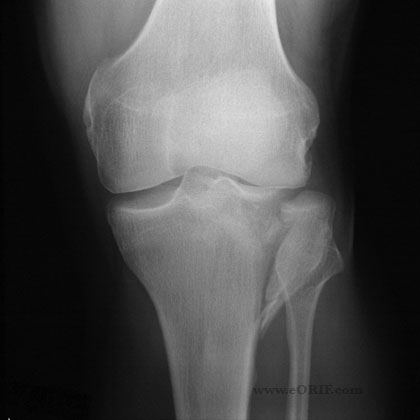
28y/o male sustained a displaced Schatzker Type II tibial plateau fracture sliding into home plate while playing softball. A/P injury view shown. |
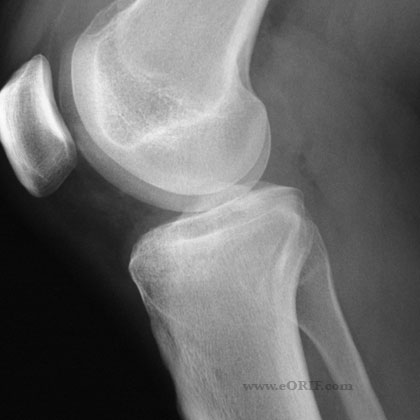 |
Lateral injury view. |
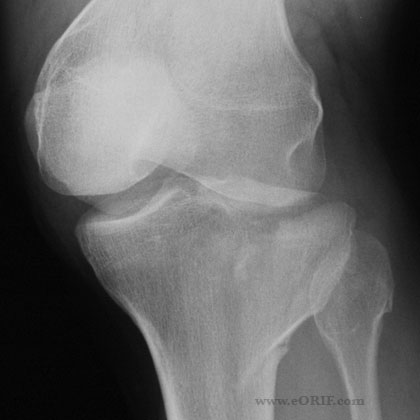 |
Oblique injury view. |
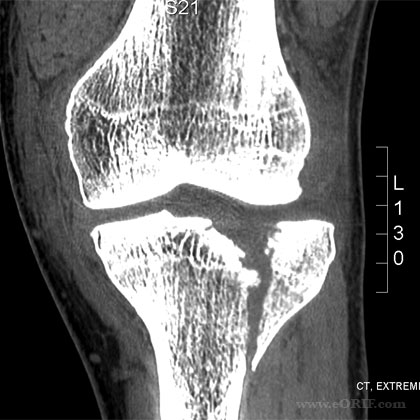 |
Coronal CT (non in traction) better shows the depressed medial aspect of the lateral tibial plateau. |
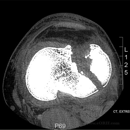 |
Axial CT (not in traction).
Knee arthroscopy was performed first. The lateral meniscus was displaced into the notch in a bucket-handle fashion with the majority of the meniscus avulsed from its capsular origin. The fracture was approach using a lateral parapatellar incision. See ORIF Technique. Reduction was obtained with longitudinal traction. The medial aspcet of the plateau was tamped back into place. The meniscus was repaired with 3 horitzontal mattress sutures. A Zimmer periarticular locking plate was selected. Two non-locking lag screws were placed proximally first. The distal screw holes filled from seperate lateral incision. |
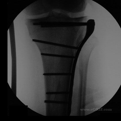 |
Intraoperative A/P view. |
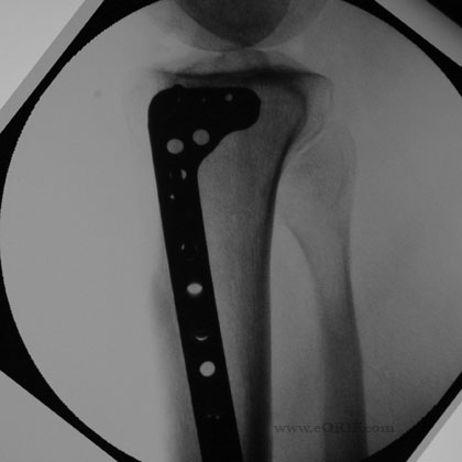 |
Intraoperative lateral view. |
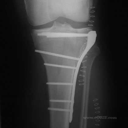 |
Post operative A/P view. |
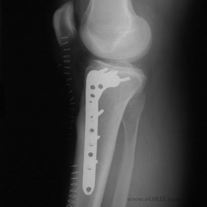 |
Post operative lateral view. |
